Compact Bone Diagram Pearson : Https Www Nlsd K12 Oh Us Userfiles 357 Classes 23426 Ch06 Lecture Pdf Id 171213. Martini, pearson education 2006, chapter 6. Compact bone is dense so that it can withstand compressive forces, while spongy (cancellous) bone has open spaces and. Learn vocabulary, terms, and more with flashcards, games, and other study tools. Most bones contain compact and spongy osseous tissue, but their distribution and concentration vary based on the bone's overall function. The following points highlight the ten main varieties of connective tissues of human body.
The walls of the diaphysis are composed of dense and hard compact bone. The cells of compact bone, which is also called cortical bone, appear to be tightly packed into a solid mass. Are the opposite of avulsion fractures. A case study on bone tissue structure and repair. Compact bone diagram pearson :
Microscopic Anatomy Of Bone Ppt Download from slideplayer.com Compact bone is the denser, stronger of the two types of bone tissue ( (figure) ). The differences between compact and spongy bone are best explored via their histology. The circulatory supply to a mature bone. A diagram of the anatomy of a bone, showing the compact bone. The two layers of compact bone and the interior spongy bone work together to protect the internal organs. Slide 5.50 each shoulder girdle attaches to the axial skeleton at only one point loose attachment of the scapula allows it to slide back and forth against the thorax as muscles act It is also called osseous tissue or cortical bone and it provides structure and support for an organism as part of its skeleton, in addition to being a location for the storage of minerals like calcium.about 80% of the weight of the human skeleton comes from. (2) axial skeleton appendicular skeleton d c c d e a b *p38751a01528* 15 turn over (c) explain the function of the joint at the shoulder.
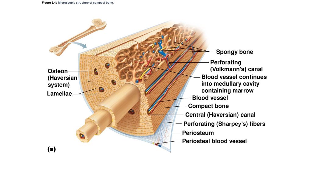
Match the following labels to the proper locations on this figure of compact bone.
A metaphysis separates the diaphysis and epiphysis at each end of the shaft. Spongy bone compact bone medullary cavity posterior view sectional view has a diaphysis (shaft) with walls of compact bone and epiphyses (ends) filled with spongy bone. Add to favorites 25 favs. Match the following labels to the proper locations on this figure of compact bone. Students will choose the term that best completes each statement about bones in the human body.
It is also called osseous tissue or cortical bone and it provides structure and support for an organism as part of its skeleton, in addition to being a location for the storage of minerals like calcium.about 80% of the weight of the human skeleton comes from.
Complete the table to show where the bones labelled in the diagram belong. In long bones, as you move from the outer cortical compact bone to the inner medullary cavity, the bone transitions to spongy bone. Clinical case study clinical case study: Compact bone, also called cortical bone, dense bone in which the bony matrix is solidly filled with organic ground substance and inorganic salts, leaving only tiny spaces (lacunae) that contain the osteocytes, or bone cells.compact bone makes up 80 percent of the human skeleton; Label the following components in the diagram of bone structure.
Http Www2 Estrellamountain Edu Faculty Hoffman Commonfiles Bio160 05theskeletalsystem 1 Pdf from Most bones contain compact and spongy osseous tissue, but their distribution and concentration vary based on the bone's overall function. Terms in this set (8) spongy bone (contains red marrow) compact bone (has osteons) osteon. This activity contains 3 questions. The hollow region in the diaphysis is called the medullary cavity, which is filled with yellow marrow. A case study on bone tissue structure and repair. Spongy bone compact bone medullary cavity posterior view sectional view has a diaphysis (shaft) with walls of compact bone and epiphyses (ends) filled with spongy bone. Evaluate your students' knowledge of the human skeletal system with this science printable. As its name indicates, spongy bone has spaces in
It can be found under the periosteum and in the diaphyses of long bones, where it provides support and protection.
The structure of a long bone, showing amongst other things the location of compact and spongy bone. On the lines below, describe the difference between spongy bone and compact bone. The walls of the diaphysis are composed of dense and hard compact bone. The diaphysis is the tubular shaft that runs between the proximal and distal ends of the bone. Complete the table to show where the bones labelled in the diagram belong. Compact bone is the denser, stronger of the two types of bone tissue ( link ). Compact bone, also called cortical bone, is the hard, stiff, smooth, thin, white bone tissue that surrounds all bones in the human body. The series of diagrams below represent the microscopic structure of compact bone tissue. Start studying osteon model, bone / osteon model. How bones react to stress. Compact bone tends to be found towards the outside of bones,. Draw a diagram of a section through a long bone. Its repeated pattern is arranged in concentric layers of solid bone tissue.
If the outer layer of a cranial bone fractures, the brain is still protected by the intact inner layer compact bone diagram. Most bones contain compact and spongy osseous tissue, but their distribution and concentration vary based on the bone's overall function.
Share :
Post a Comment
for "Compact Bone Diagram Pearson : Https Www Nlsd K12 Oh Us Userfiles 357 Classes 23426 Ch06 Lecture Pdf Id 171213"


Post a Comment for "Compact Bone Diagram Pearson : Https Www Nlsd K12 Oh Us Userfiles 357 Classes 23426 Ch06 Lecture Pdf Id 171213"